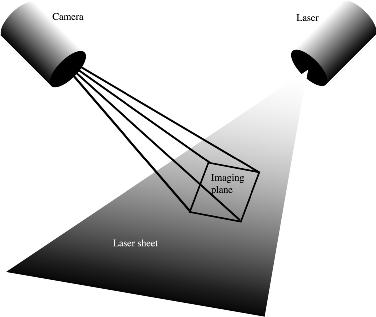
Working with Jules Jaffe of Scripps, we have developed a novel two-dimensional imaging fluorometer. This system uses a sheet of laser light to induce chlorophyll a fluorescence of phytoplankton, which is imaged using a sensitive CCD camera. The laser sheet is angled downward at 45 degrees to extend below the platform as it sinks. This permits imaging of plankton that are undisturbed by the instrument itself.
In July of 2001 we deployed a second-generation imaging fluorometer off San Diego, CA, on an untethered free-falling platform. We could regulate the buoyancy to set the fall speed, typically 3-15 cm/s. Below are some sample images obtained from the system.
One striking feature of the images is the discrete nature of the phytoplankton. Rather than forming a continuous sheet of fluorescent material, we clearly see the individual cells, with considerable empty space between them. The camera system (imaging 35 x 35 cm with 300 micron resolution) can distinguish between single dinoflagellates and diatom chains.
The images below have not been processed: they must have the camera's dark current removed, and then be corrected for the pattern of excitation illumination. Once corrected they can be analyzed for the details of the microscale distribution patterns of the plankton.
 Subsurface chlorophyll maximum layer
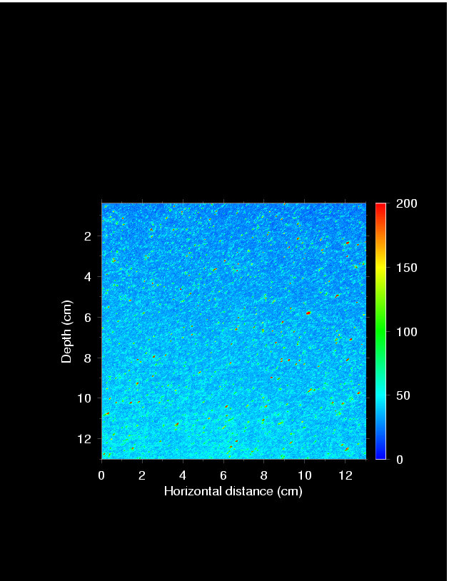
at 35 m depth. This image is 13x13 cm and shows a strong vertical gradient of the background chlorophyll (cells smaller than about 5 microns), as well as increased numbers of large, intensely fluorescent cells. Such vertical gradients are common in the images.
Subsurface chlorophyll maximum layer
at 35 m depth. This image is 13x13 cm and shows a strong vertical gradient of the background chlorophyll (cells smaller than about 5 microns), as well as increased numbers of large, intensely fluorescent cells. Such vertical gradients are common in the images. Dinoflagellates
at 14 m depth. Note the individual cells.
Diatom chains
at 35 m depth in the fluorescence maximum. This image is not smeared - note the variety of orientations of the cells. Our bottle sampling underpredicted the average length of the chains, as they broke being poured from the bottle.
Side-scattered light
recorded by the camera with the chlorophyll filter removed. Compare this image with those from above: there are many more particles, and more larger particles than appear in the fluorescence images.
Side-scattered image of particles being mixed by the instrument platform as it heaved up and down when attached to the ship. Notice the small-scale flow stuctures within the image.
Dinoflagellates
at 14 m depth. Note the individual cells.
Diatom chains
at 35 m depth in the fluorescence maximum. This image is not smeared - note the variety of orientations of the cells. Our bottle sampling underpredicted the average length of the chains, as they broke being poured from the bottle.
Side-scattered light
recorded by the camera with the chlorophyll filter removed. Compare this image with those from above: there are many more particles, and more larger particles than appear in the fluorescence images.
Side-scattered image of particles being mixed by the instrument platform as it heaved up and down when attached to the ship. Notice the small-scale flow stuctures within the image.E-mail below for a reprint